

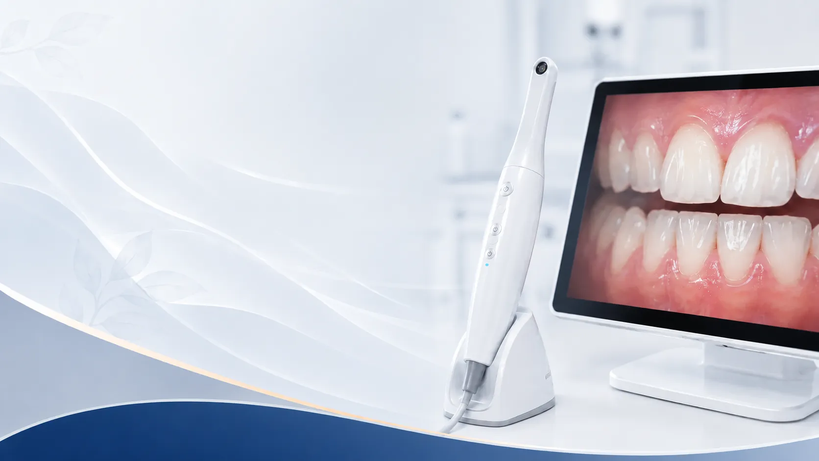
See the Area Being Discussed Instead of Relying Only on a Description
Morton Family Dentistry uses small intraoral cameras to make dental findings easier to show, document, and understand.
Many dental problems occur in places patients cannot see in a bathroom mirror. An intraoral camera lets the Morton dental team take enlarged photographs inside the mouth and display them during the visit.
Seeing the actual tooth or gum area can make a recommendation easier to understand. It also gives the patient a clearer basis for questions before deciding on treatment.
Details the Camera May Capture
- A chipped edge or visible crack
- Wear around a filling, crown, or bridge
- Plaque, tartar, or inflammation near the gumline
- A suspicious spot on the visible tooth surface
- Changes that should be compared at a future appointment
Photography and X-Rays Show Different Information
The camera records what can be seen on the surface. X-rays reveal structures beneath enamel, between teeth, and within bone. The dentist may use one or both depending on the question being investigated.
Useful for Documentation
Images can become part of the dental record, making it easier to monitor a worn area, compare a restoration over time, or document the condition of a tooth before treatment.
No Radiation and No Recovery
The camera is small, and the process is similar to moving a dental mirror around the mouth. It does not use radiation and does not require numbing.
Choose a Dental Office That Shows Its Findings
Morton Family Dentistry uses visual tools to support clear explanations and informed decisions during routine and problem-focused visits.
Frequently Asked Questions
Does the camera hurt?
No. It is a non-invasive imaging tool, although patients with a sensitive gag reflex can let the team know so positioning can be adjusted.
Can I receive a copy of an image?
Ask the dental team. Images are stored as part of the clinical record and may be available for treatment coordination or patient education.